
How a Haematology Analyser Works: From Tube to FBC Report
What happens inside an FBC machine in the minute after you load a tube: how cells are counted and sized, how a 5-part analyser tells white cells apart, how haemoglobin is measured, and which numbers on the report are calculated rather than measured.
One sample, up to three measuring channels
The analyser aspirates a few microlitres of well-mixed EDTA blood and splits it. One portion is diluted heavily in an isotonic diluent so red cells and platelets can be counted. Another is mixed with a lyse reagent that destroys the red cells, so white cells can be counted and the freed haemoglobin measured. A 5-part analyser adds an optical channel in which a laser classifies each white cell. In one widely used design, blood for the red-cell and platelet count is diluted about 6,000-fold and blood for the white-cell count about 250-fold.
| Channel | Reagent added | What is measured | Method |
|---|---|---|---|
| RBC / PLT | Diluent only | Red cells, platelets and their volumes | Impedance (Coulter principle) |
| WBC / HGB | Diluent and lyse | White-cell count, 3-part sizing and haemoglobin | Impedance for cells; photometry for haemoglobin |
| Optical (5-part analysers) | Lyse plus a stain or fluorescent dye | Five white-cell types; on some models NRBC, reticulocytes and immature granulocytes | Laser light scatter and fluorescence (flow cytometry) |
The impedance (Coulter) principle
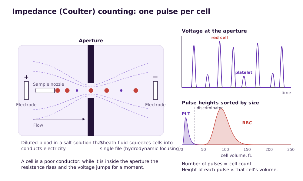
Wallace Coulter’s method, patented in 1953 and still the basis of most FBC machines, rests on one fact: blood cells are poor electrical conductors. Suspend them in a salt solution, draw it through a tiny aperture with an electrode on each side, and every cell that passes briefly raises the resistance of the aperture. Under a constant current that shows up as a voltage pulse. The number of pulses is the cell count; the height of each pulse is proportional to that cell’s volume.
Counting
Pulses are counted in a fixed volume of diluted sample and converted back to cells per litre of blood using the dilution factor.
Sizing
Pulse heights are sorted into a size histogram in femtolitres (fL). Red cells cluster around 80–96 fL in healthy adults and platelets far below; a discriminator between the two decides which pulses are counted as which.
Reading the histogramsCoincidence
Two cells crossing the aperture together make one large pulse. Heavy dilution and hydrodynamic focusing keep this rare, and the analyser corrects for what remains.
From Model A to the clinic analyser
Coulter’s Model A was the first commercial haematology analyser. By 1977 the Model S Plus added a three-part white-cell differential by size, the approach compact 3-part clinic analysers still use.
Read next Blood cell counters compared
Hydrodynamic focusing: one cell at a time, through the centre
A cell that drifts through the edge of the aperture produces a distorted pulse and can be sized wrongly. Modern analysers inject the sample through a fine nozzle into a stream of sheath fluid, which narrows it into a single-file thread through the centre of the aperture. The FDA’s decision summary for Sysmex’s compact XW-100, for example, describes direct current with hydrodynamic focusing for every parameter except haemoglobin, which is measured photometrically.
- Fewer coincidence events, so counts stay accurate at high cell numbers.
- More consistent pulse heights, so a tighter, more reliable MCV and RDW.
- Cells pass the sensing zone once, so they are not counted twice.
Optical flow cytometry: how a 5-part analyser sorts white cells
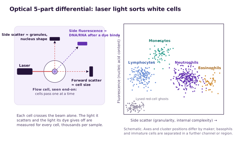
Size alone separates white cells into three groups. To report neutrophils, lymphocytes, monocytes, eosinophils and basophils separately, a 5-part analyser passes each white cell through a laser beam and measures the light it scatters. Forward scatter rises with cell size. Side scatter, measured at about 90°, rises with internal complexity such as granules and a lobed nucleus. Where a fluorescent dye is added, side fluorescence shows how much nucleic acid the cell holds. Sysmex’s XN-series, for example, reads forward scatter, side scatter and side fluorescence from a 633 nm laser.
Other makers, other signals
Not every 5-part analyser uses fluorescence. Beckman Coulter’s DxH analysers, for instance, combine cell volume, conductivity (opacity) and light scatter for each cell.
When clusters run together
Abnormal cells such as blasts or immature granulocytes fall between or outside the normal clusters. The analyser cannot name them, so it flags the result for a blood film.
What the flags mean| Cell | Where it falls | Why |
|---|---|---|
| Lymphocytes | Low side scatter | Small cells with few granules |
| Monocytes | Moderate scatter, high fluorescence | Larger cells with a folded nucleus and more nucleic acid than neutrophils |
| Neutrophils | High side scatter | Granular cytoplasm and a lobed nucleus |
| Eosinophils | Highest side scatter | Large, dense granules |
| Basophils and immature cells | A further channel or region, depending on the maker | They overlap other cells in the main plot |
Haemoglobin: photometry, now without cyanide
The lyse reagent breaks open the red cells, and the freed haemoglobin is converted to a single stable coloured compound whose absorbance a photometer reads. The reference method converts it to cyanmethaemoglobin, but cyanide waste is a hazard, so most analysers now use cyanide-free reagents. The best known, sodium lauryl sulfate (SLS) haemoglobin, was published by Oshiro and Takenaka in 1982 and is used by several analyser makers.
- A blank reading of the reagents is taken in each cycle, so the analyser measures only what the blood adds.
- Anything that makes the solution cloudy, such as lipaemia or a very high white-cell count, can raise the haemoglobin reading, and with it MCH and MCHC.
- Because the lyse is shared, too little of it leaves red cells unlysed, which raises both the white-cell count and the haemoglobin.
Measured or calculated? Reading the report with that in mind
Only some lines on an FBC report are measured directly. The rest are calculated from them, so an error in one measured value spreads into every index built on it. That is why an impossible MCHC is such a useful warning sign.
| Parameter | How it is obtained | Formula or source |
|---|---|---|
| WBC, RBC, PLT | Measured (counted) | Impedance pulses, or optical events |
| HGB | Measured | Photometry after lysis |
| MCV | Measured on impedance analysers | Mean volume of all counted red-cell pulses |
| HCT | Calculated | RBC × MCV |
| MCH | Calculated | HGB ÷ RBC |
| MCHC | Calculated | HGB ÷ HCT |
| RDW | From the RBC histogram | Spread of red-cell volumes |
| MPV, PDW | From the PLT histogram | Mean and spread of platelet volumes |
| PCT (plateletcrit) | Calculated | PLT × MPV |
| Differential (% and #) | Measured (classified) | Size on a 3-part; scatter and fluorescence on a 5-part |
Read next How to read FBC resultsFBC normal ranges in Malaysia
What is optimal: sample age, mixing and QC timing
The analyser is only as good as the tube it is given and the controls run before it. These habits give the most reliable result.
| Step | Optimal practice | Why |
|---|---|---|
| Tube | K2 or K3 EDTA, filled to the line | An under-filled tube leaves excess EDTA, which shrinks red cells and lowers HCT and MCV |
| Mixing at collection | Invert gently 8–10 times straight away | The count BD gives for EDTA tubes; small clots ruin the platelet count |
| Mixing before analysis | Re-mix by gentle inversion just before aspiration | Red cells settle on standing; an unmixed sample reads falsely low or high |
| Sample age | Run within a few hours; for a differential, within 8 hours at room temperature | Boule states its M32 needs samples under 8 hours old for a correct white-cell differential |
| If there is a delay | Refrigerate at 4 °C and run within 12 hours | In a 48-hour study, differential counts changed significantly after 12 hours at 23 °C, while 4 °C kept the full count and differential stable for 12 hours |
| Controls | Two levels before patient samples each testing day, and after every reagent lot change, maintenance or service | Catches drift before it reaches a patient result; follow your maker’s interval where it is stricter |
| Background | A blank at start-up, within the maker’s limit | A high background adds false counts, most visibly to platelets |
Read next Daily QC and EQAHandling the EDTA sample
Related guides
Hematology Analyser supply near you
Written quotes for clinics across Malaysia, handled from Batu Caves, Kota Kinabalu and Kuching.
- Hematology Analyser Kuala LumpurKuala Lumpur
- Hematology Analyser SelangorSelangor
- Hematology Analyser Petaling JayaSelangor
- Hematology Analyser Shah AlamSelangor
- Hematology Analyser KlangSelangor
- Hematology Analyser SerembanNegeri Sembilan
- Hematology Analyser MelakaMelaka
- Hematology Analyser Johor BahruJohor
Sources and further reading
- Graham MD. The Coulter Principle: a history. Cytometry Part A, 2022
- Developments of conventional and microfluidic flow cytometry enabling high-throughput characterization of single cells (review, 2022)
- Oshiro I, Takenaka T. New method for hemoglobin determination by using sodium lauryl sulfate (SLS). Clinical Biochemistry, 1982
- US FDA decision summary K210346: XW-100 Automated Hematology Analyzer for CLIA-waived use
- Boule Diagnostics. Understanding histograms: Medonic M32 white paper
- Stability of hematological analytes during 48 hours storage at three temperatures using a Cell-Dyn analyzer (2021)
Frequently asked questions
What principle does a haematology analyser use?
Is the haematocrit measured or calculated?
Why does a 5-part analyser need a laser?
How long after collection can an FBC be run?
Who invented the blood cell counter?
Get a written quotation
Tell us your clinic or lab type, daily workload and location. We match suitable models and send a written quotation that states who handles delivery, installation and training, and what each costs.
010-905 2891 · Mon–Sat, 9:00 am – 6:00 pm